Q.8 Describe structure and mechanism of function of human eye.
Answer:
Human Eye
Location
Our eyes are located in small portions of skull known as the orbits or eye sockets.
External Structure of Eye
(i) Eyelids
Functions
Eyelids wipe eyes and prevent dehydration. They spread tears on eyes which contain substances for fighting bacterial infections.
(ii) Eyelashes
Function
Eyelashes prevent fine particles from entering eye.
Internal structure of eye
The internal structure of eye can be divided into three main layers;
(i) Outer layer
The outer layer of eyeball consists of sclera and cornea.
(a) Sclera
It consists of dense connective tissue.
Functions
Sclera gives eye most of its white colour, It protects the inner components of eye.
It maintains the shape of eye.
(b) Cornea
In the front, sclera forms the transparent cornea.
Function
Cornea admits light to the interior of eye and bends light rays so that they can be brought to a focus. (ii) Middle Layer Choroid
The middle layer is called choroid.
Structure
It contains blood vessels.
Function
It gives the inner eye a dark colour. The dark colour prevents disruptive reflections – within eye.
IRIS
Behind cornea, choroid bends to form a muscular ring called iris.
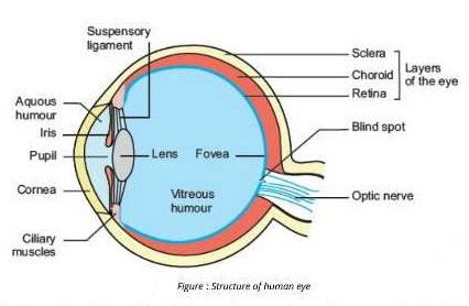
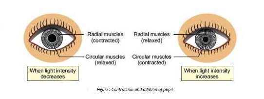
Pupil
There is a round hole, called pupil, in the centre of iris. After striking the cornea, light passes through the pupil.
Size
The size of pupil is adjusted by the muscles of iris.
Function
Pupil constricts in bright light when the circular muscles of iris contract Pupil dilates in dim light when the radial muscles of iris contract.
Lens
Behind iris, there is a convex lens.
Function
It focuses light on the retina.
Ciliary muscles and Suspensory Ligaments
Lens is attached to ciliary muscles of eye via/through a ring of suspensory ligament.
Function
To see an object far clearly that is away, ciliary muscles are relaxed and lens becomes less convex. When ciliary muscles contract, lens becomes more convex and round.
(iii)INNER LAYER-RETINA
The inner layer is sensory and is called retina. It contains the photosensitive cells called rods and cones and associated neurons.
Rods
These are sensitive to dim light.
Cones
These are sensitive to bright light and they distinguish different colours.
Points on Retina
Retina has two points, fovea and optic disc.
Fovea
Fovea is a dip in retina directly opposite to lens and is densely packed with cone cells.
Function
It is largely responsible for colour vision and sharpness.
OPTIC DISC
Optic disc is a point on retina where the optic nerve enters retina.
Blind spot
There are no rods and cones at this point, that is why it is also referred to as the blind spot. Chambers
The iris divides the cavity of eye into two chambers.
Anterior chamber
The anterior chamber is in front of iris i.e. between cornea and iris.
Aqueous humour
The anterior chamber contains a clear fluid known as aqueous humour.
Posterior chamber
The posterior chamber is between iris and retina.
VITREOUS HUMOUR
The posterior chamber contains a jelly – like fluid known as vitreous humour.
Function
It helps to maintain the shape of eye and suspends the delicate lens.
MECHANISM OF VISION
Light from objects enters eye and is refracted when it passes through cornea, aqueous humour, lens and vitreous humour. Lens also focuses light on retina. As a result, the image falls on retina. Rods and cones generate nerve impulses in the optic nerve. These impulses are carried to the brain, which makes the sensation of the vision.
![]()